BioMEMS and Medical Devices
The microfluidics team at the Ali Khademhosseini laboratory has developed a wide variety of platforms for gradient microfluidics, microassay microfluidics, and high-throughput screening (HTS) stem cell microfluidics. Platform development includes both experiment and theory. These approaches are beneficial for developing practical biomimetic microsystems that can enhance the function of organs and their future regeneration.
Recent projects by the microfluidics team include:
a) Gradient microfluidics for fast generation of molecule, particle and cell gradients
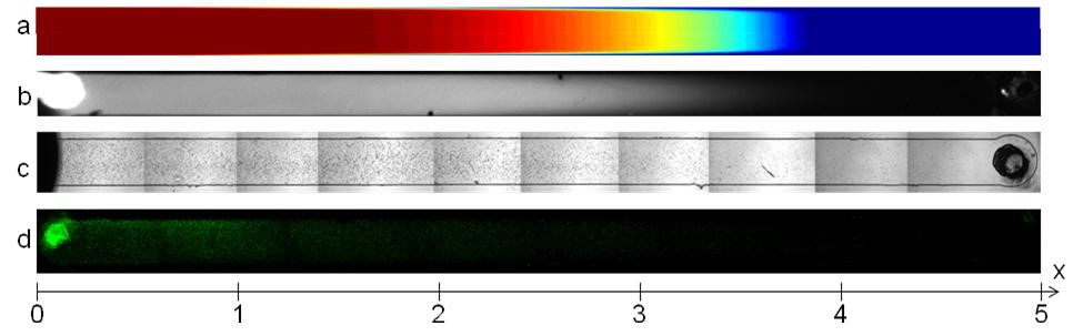
Figure 1: Gradients generated by rapid flow and flow reversal. (a) simulation of chemical gradient, and experiments (b) FITC-dextran, (c) cells and (d) 5 ?m beads. [Y. Du, M. J. Hancock, J. L. Villa-Uribe, D. Cropek, and A. Khademhosseini]
b) Stem cell microfluidics (with pneumatic mechanical compression as stimuli)
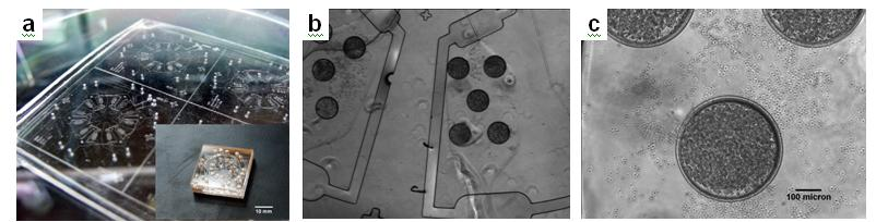
Figure2: (a) Photograph of a micro cell chip integrated with various sizes of microwells and microvalving systems. Scale bar is 10 mm (b) Phase contrast images of the EB formation after cell loading in 300 ?m-diameter microwells. (c) Phase image of individual microwells with higher magnification. [W. Y. Sim, W. G. Lee, Y.S. Sik, and A. Khademhosseini]
c) Stem cell microfluidics (for high-throughput screening of stem cell microenvironments)
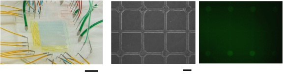
Figure3: (a) Photograph of a visualized microfluidic screening device and its function. Scale bar is 10 mm. This device was used to characterize embryoid body (EB) formation of Gsc-embryonic stem cells and 5-day culture of the EBs within the microwells of the microdevice. (b) Phase contrast and fluorescent images of 5-day cultured EBs. Scale bar is 300 ?m. [W. G. Lee, W. Y. Sim, B. G. Chung, J. W. Hong, and A. Khademhosseini]